Gastric Tube For Nutrition: Procedure And Advantages
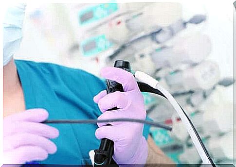
Gastrostomy is a surgical procedure in which a feeding tube is placed. This surgery is performed through a minimally invasive operation, with endoscopic techniques. For this reason it is also known as percutaneous endoscopic gastrostomy, or simply PEG.
The introduction of the probe into the stomach allows the food to arrive safely in subjects who do not have a normal digestive function.
It is very common to use the gastric tube when the patient suffers from dysphagia (typical of amyotrophic lateral sclerosis or ALS), or difficulty in swallowing the food bolus. It is also common in patients who have suffered a stroke or other type of neurological damage.
When comparing percutaneous endoscopic gastrostomy with the nasogastric tube, which is placed from the nose to the stomach, the first option has many more advantages. First, the significant reduction of complications derived from the probe itself. For example, infections or injuries associated with the pharynx, caused by friction with the tube.
By skipping most of the digestive tract path, the PEG is also safer for the patient. There is also an improvement in the psychological sphere, because the gastric tube is a much more discreet device than the nasogastric tube. This aspect is very important especially for young patients, who are concerned about the aesthetic limitation of the classic tube.
All the steps to apply the gavage
For the patient, the gastric tube offers a high number of advantages, both medical and aesthetic, compared to the nasogastric alternative. In the following lines we see how to proceed for its application.
1. How does it fit?

As we just explained, this is a pretty straightforward process. To explain the individual steps that make up gavage insertion, we will refer to the information provided by the American Society of Gastrointestinal Endoscopy.
First, an intravenous solution is administered to sedate the patient, in an endoscopy room. The most appropriate region of the abdomen is located according to the results of endoscopy, defined by the US National Library of Endoscopy as a tool for observing the inside of the body. Then we proceed to disinfect the area.
Subsequently, the area is anesthetized with a local anesthetic and an incision is made on the abdominal wall. It is a small cut, 1 cm more or less, so postoperative recovery is quick and with little risk.
Immediately thereafter, a trocar is introduced to the stomach, using the endoscope cable as a guide. The trocar is a kind of metal cannula capable of penetrating the stomach wall and reaching the inside of the organ.
Finally, the endoscope is withdrawn by rewinding the cable to the patient’s mouth and the gastric tube is positioned, which will come out of the abdominal incision. For added safety, manufacturers include a balloon that inflates to prevent the probe from moving. In this way it remains stationary and can be closed comfortably.
When should the gastric tube be replaced?
PEG, as we have seen, is a fairly safe process. However, since these are patients who need prolonged enteral feeding , some complications may occur and the tube may need to be removed.
The most common complication is infection, although bleeding is also possible, as reported by the MedlinePlus medical encyclopedia .
Regarding the infection, the latter can originate in the gastrostomy orifice on the abdominal wall or it can be caused by the tube itself. In any case, it is mandatory to remove it and start antibiotic therapy to eliminate the infection, before a serious picture of septicemia occurs.
How is it replaced?
To remove the gastric tube, you must first sterilize and disinfect the area around the gastrostomy and stop feeding, so that the tube remains clean.
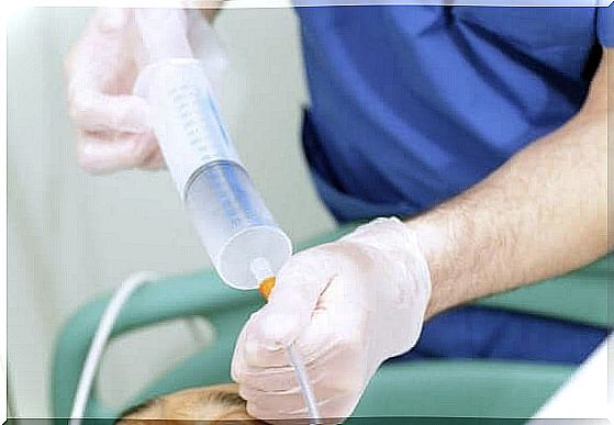
Next, the balloon of the new probe is checked for proper functioning. To check this, sterile water is introduced into the valve to inflate the balloon. If it inflates correctly and there are no leaks, then it can be used.
The area around the orifice of the gastric tube is carefully cleaned and the balloon that prevented it from coming out is deflated. When the probe is not working and is not fixed, it can be removed by pulling with one hand, while pressing the abdomen with the other.
Finally, the new gastric tube is introduced through the same gastrostomy orifice. The ideal situation is one in which the tube forms a right angle to the abdomen to facilitate insertion.
Finally, the balloon is inflated to secure the probe and gently pulled until it rests against the gastric wall. Of course, you should always disinfect once the replacement is complete, to prevent possible infection.
A force majeure decision
In conclusion, the gavage is used in cases of force majeure, when the patient, for a variety of reasons (for example, a stroke), is unable to feed on his own.
While inserting the probe is a simple procedure, it is still a surgical operation and, as a result, complications may arise. Naturally, the doctor will evaluate the possible need to apply it, based on the needs of each individual patient.